RETINA
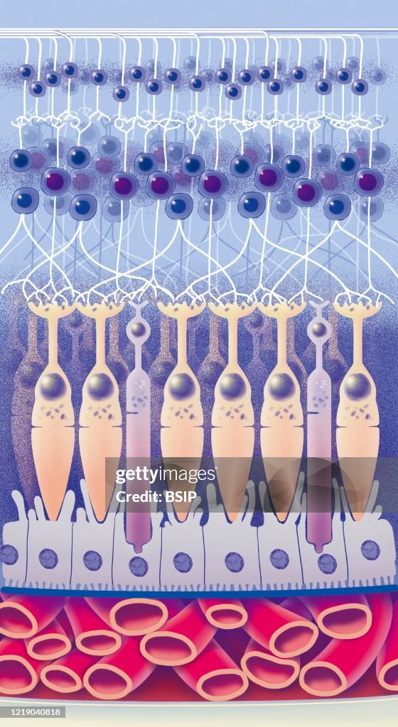
Zoom on the retina and its different layers. From the bottom up the scoteroid (yellowish-white), the choroid consisting of vessels, separated from the retina by Bruch's membrane (blue). The retina consists of pigment epithelium (light purple) cones and rods (orange), an inner granular layer with horizontal cells, bipolar and amacrine, a layer of ganglion cells and a layer of nerve fibers (axons ganglion cells). (Photo by: BSIP/Education Images/Universal Images Group via Getty Images)

PURCHASE A LICENSE
How can I use this image?
kr 2,500.00
NOK
Getty ImagesRETINA, News Photo RETINA Get premium, high resolution news photos at Getty ImagesProduct #:1219040818
 RETINA Get premium, high resolution news photos at Getty ImagesProduct #:1219040818
kr4,000kr950
Getty Images
In stockPlease note: images depicting historical events may contain themes, or have descriptions, that do not reflect current understanding. They are provided in a historical context. Learn more.
DETAILS
Restrictions:
Contact your local office for all commercial or promotional uses.
Credit:
Editorial #:
1219040818
Collection:
Universal Images Group
Date created:
November 30, 2019
Upload date:
License type:
Release info:
Not released. More information
Source:
Universal Images Group Editorial
Object name:
941_28_bsip_015927_140.jpg
Max file size:
2789 x 5100 px (9.30 x 17.00 in) - 300 dpi - 4 MB