Small intestine, LM - stock photo
Small intestine. Light micrograph (LM) of a section through the finger-like projections (villi) of the duodenum, the uppermost part of the small intestine. These increase the surface area for the absorption of food. Within the columnar epithelium of the outer surface (blue) are goblet cells (red), which secrete mucus to lubricate food and prevent self-digestion. The lamina propria (central core, blue) contains the blood supply that transports the products of digestion. Magnification: x500 when printed at 10 centimetres wide.

Get this image in a variety of framing options at Photos.com.
PURCHASE A LICENSE
All Royalty-Free licenses include global use rights, comprehensive protection, simple pricing with volume discounts available
kr 2,500.00
NOK
Getty ImagesSmall Intestine Lm High-Res Stock Photo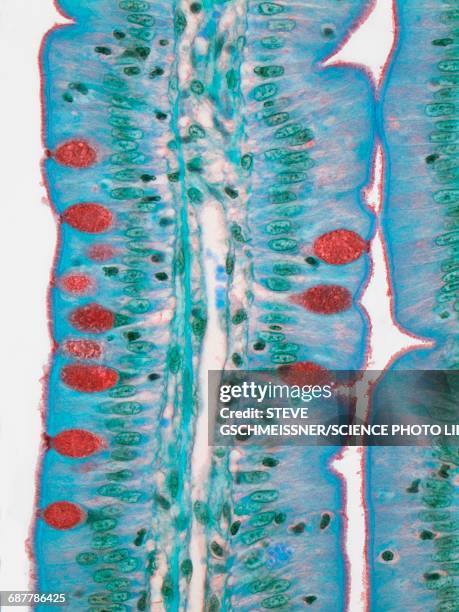 Download premium, authentic Small intestine, LM stock photos from Getty Images. Explore similar high-resolution stock photos in our expansive visual catalogue.Product #:687786425
Download premium, authentic Small intestine, LM stock photos from Getty Images. Explore similar high-resolution stock photos in our expansive visual catalogue.Product #:687786425kr2,500kr300
Getty Images
In stockDETAILS
Creative #:
687786425
License type:
Collection:
Science Photo Library
Max file size:
3619 x 4829 px (12.06 x 16.10 in) - 300 dpi - 5 MB
Upload date:
Release info:
No release required
Categories:
- Goblet Cell,
- Microscope,
- Villus,
- Alternative Medicine,
- Anatomy,
- Biology,
- Built Structure,
- Color Image,
- Columnar Epithelium,
- Digestive System,
- Duodenum,
- Healthcare And Medicine,
- Healthy Lifestyle,
- Histology,
- Human Tissue,
- Ileum,
- Intestine,
- Jejunum,
- Lamina Propria,
- Light Micrograph,
- Magnification,
- Membrane,
- Microbiology,
- Microvillus,
- Mucus,
- No People,
- Photography,
- Science,
- Scientific Imaging Technique,
- Secreting,
- Secretory Cell,
- Small Intestine,
- Tissue - Anatomy,
- Vertical,