Dividing cancer cell, fluorescent microscopy - HD stock video
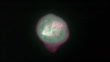
Timelapse fluorescence microscopy video of a dividing cancer cell in 3D tissue culture. The total length of the sequence is 3 hours. The cell is expressing a fluorescently-tagged version of a protein (EB1) that binds to growing microtubule ends and also localizes to the spindle poles. This highlights the highly dynamic microtubule cytoskeleton in the spindle, and also shows astral microtubules extending toward the cell periphery. The different colors represent different focal planes giving a more 3D impression. In the beginning of the video it can be seen how the duplicated centrosomes move around the cell nucleus and then form the poles on opposite ends of the mitotic spindle apparatus. Although chromosomes themselves are not labeled, they can be seen as darker shadows in the cytoplasm. The cell eventually divides apparently without all chromosomes having aligned correctly in the middle of the spindle, leading to aneuploidy, one of the hallmarks of cancer. This clip is a slowed-down version of K007/4478


PURCHASE A LICENSE
Get personalized pricing by telling us when, where, and how you want to use this asset.
DETAILS
Credit:
Creative #:
1415596608
License type:
Rights-ready
Collection:
Photolibrary Video
Max file size:
1920 x 1080 px - 399 MB
Clip length:
00:00:32:00
Upload date:
Location:
United States
Release info:
No release required
Mastered to:
QuickTime 8-bit Photo-JPEG HD 1920x1080 25p
Categories:
- Cancer Cell,
- Cancer - Illness,
- Cell Division,
- Mobile Phone,
- Scientific Experiment,
- Metastasis,
- Light Micrograph,
- Mitosis,
- Research,
- Anaphase,
- Biology,
- Bizarre,
- Black Background,
- Chromosome,
- Color Image,
- Condition,
- Cytoskeleton,
- Film - Moving Image,
- HD Format,
- Horizontal,
- Illness,
- Magnification,
- Metaphase,
- Microtubule,
- Mitotic,
- Oncology,
- Prophase,
- Repetition,
- Spindle,
- Super Slow Motion,
- Time Lapse,
- USA,
- Wireless Technology,